 Lebanese Ministry of Information
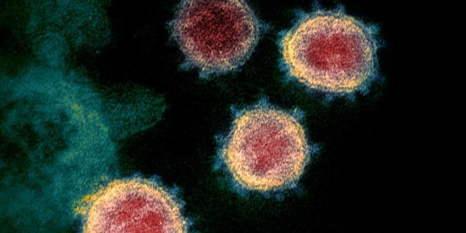
Researchers at the University of Minnesota created a 3D map of the ‘spike’ protein used to latch on to cells
New research has shed light on a crucial biological mechanism that may have helped the coronavirus to infect humans and spread rapidly around the world.
A detailed analysis of the virus’s structure shows that the club-like “spikes” that it uses to establish infections latch on to human cells about four times more strongly than those on the related Sars coronavirus, which killed hundreds of people in a 2002 epidemic.
The finding suggests that coronavirus particles that are inhaled through the nose or mouth have a high chance of attaching to cells in the upper respiratory tract, meaning that relatively few are needed for an infection to gain a foothold.
Scientists at the University of Minnesota used X-ray crystallography to create an atomic-scale 3D map of the virus’s spike protein and its corresponding partner on human cells, known as the ACE-2 receptor.
When the virus encounters a human cell, the spike proteins on its surface stick to ACE-2 receptors, if the cell possess them, and allow the virus to gain access and replicate.
“The 3D structure shows that compared to the virus that caused the 2002-2003 Sars outbreak, the new coronavirus has evolved new strategies to bind to its human receptor, resulting in tighter binding,” said Dr Fang Li, who led the US team. “The tight binding to the human receptor can help the virus infect human cells and spread among humans.”
The map of the virus will now be used by scientists to search for potential drugs that can neutralise the virus before replication has ramped up and the infection has taken hold. “If a new antibody drug can bind to those sites on the virus more strongly and frequently than the receptor, it will block the virus out of cells, making it a potentially effective treatment for viral infections,” Li said. The same sites can be used to shape work on vaccines to prevent future infections, he added.
The Guardian